Diagram Of Cartilage
Long bone diagram hyaline cartilage Cartilage sac The expressive figure: cartilage of nose
The Expressive Figure: Cartilage of Nose
Cartilage skeleton skeletal bones fibrocartilage body location elastic tissues hyaline types scientistcindy Cartilage joints hyaline bones strongest fibrocartilage myfamilyphysio slippery smoothly enables External ear – oto surgery atlas
Cartilage: types of cartilage
Tracheal cartilage diagramCartilage bone between differences structure tissue any Cartilage articular orthobullets layers basic topicCartilaginous joints joint anatomy symphysis cartilage synchondrosis epiphyseal hyaline plate pubic bones pelvis fibrocartilage hip articulation body left bone figure.
Joints cartilaginous skeleton types joint bones synchondrosis two anatomy ligaments body cartilage human learn synovial example fibrous skeletons structure theirCartilages laryngeal larynx skeleton major paired epiglottis stridor neck fig relation teachmeanatomy unpaired Cartilage bones functions 1303 osteoarthritis cartilages cartilagoCartilage histology fibrocartilage fibro.

Joints and ligaments
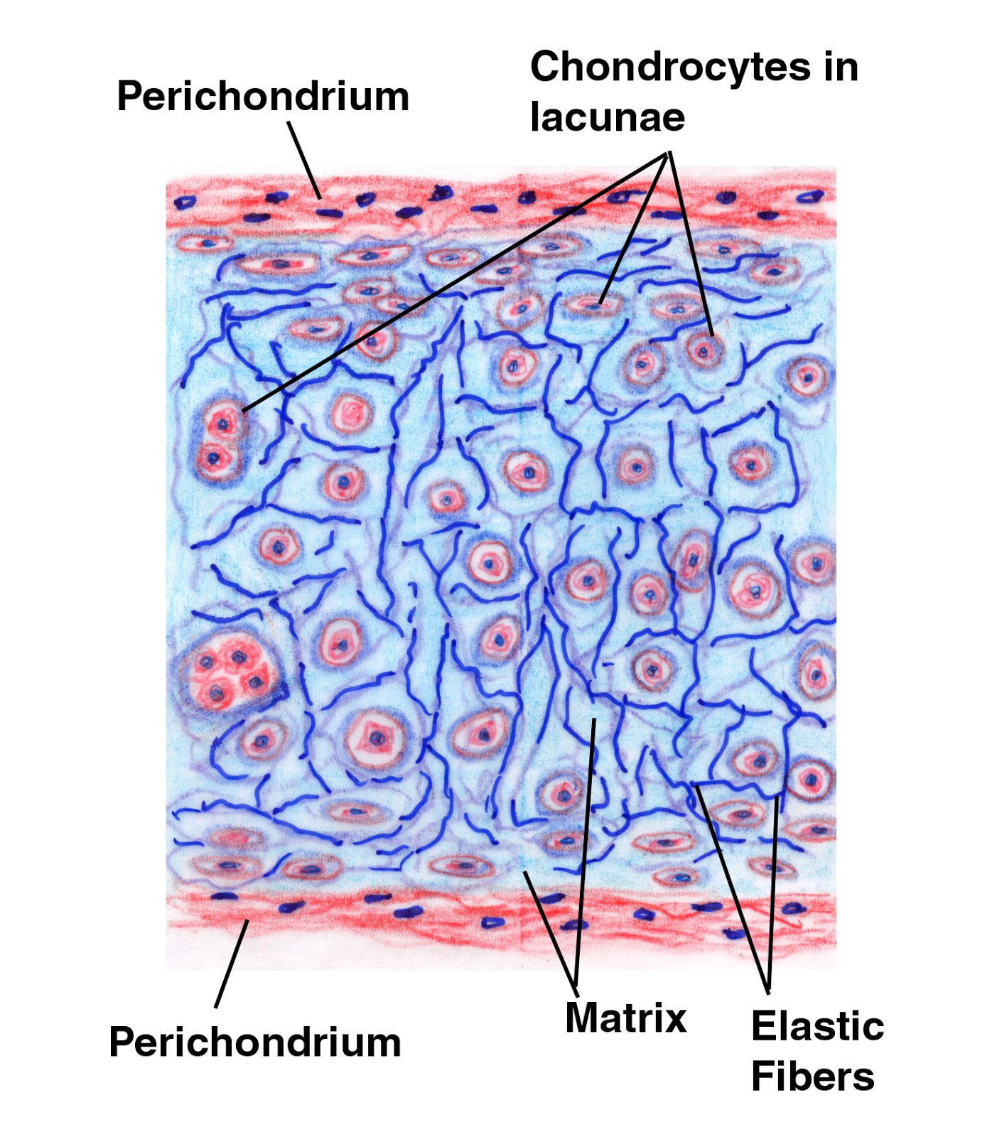
Figure 1 from articular cartilage : structure , composition , injuriesCartilage perichondrium layer chondrogenic histology inner cells anatomy vessels chondrocytes features anatomyqa Why do babies have more bones than adults?Pictures of cartilage.
Generation of cartilage from periosteum in vivo, a clear clue how toArticular cartilage Nose cartilage diagram alar anatomy base nasal valve external surgery collapse stenosis revision facial human figure interesting factsCartilage and bones.

Cartilage hyaline bone fibrous cartilaginous articular synovial
Ear cartilage anatomy auricle helix crus external incisura tragus incision skeleton surgery atlas location surgical note betweenCartilage articular composition injuries Differences between bone and cartilageHow pemf therapy accelerates articular cartilage repair ~ pemf therapy.
Cartilage hyaline membrane vessels know details clickCartilage articular repair pemf therapy Cartilage periosteum bone articular diagram histology vivo generation ireland pointsPictures of cartilage.

What is cartilage?
Hyaline cartilageThyroid cartilage anatomy, function & thyroid cartilage fracture Cartilage bone joint part ligament medical synovial section cross between membrane typical tissues fluid showing hyaline types bones tissue articularCartilage articular tissue compression diagram injury end jose biology connective intercellular pliable resists solid material does.
Histology image: cartilageCartilaginous joints · anatomy and physiology Trachea anatomy trakea cartilage organ tracheal dan keterangannya pernapasan esophagus materikimia functi healthjadeCartilage joints.

Cartilage types biology function definition
Cartilage healthiackCartilage hyaline boneandspine connective collagen Laryngeal cartilages.::jose biology b::.: .::vocabulary of concepts of ch.33 animal tissue::..
Chondrocytes cartilage difference between vs bone diagram cells called figureThyroid cartilage laryngeal cricoid membrane ligaments fracture Bone vs. cartilageCartilage types, structure and function.

Cartilage hyaline bones elastic skeletal body bone human system tissue types joints fibrocartilage location locations anatomy their organs where found
Bone fibrocartilage cartilage histology anatomy endochondral symphysis pubic marrow formation zone physiology tissue between studyIowa research focused on cartilage repair Cartilage articular tissue engineering schematic scaffold cell frontiersin biocompatible applications chondrocytes depiction joint fbioeCartilage facty 7activestudio.
Cartilage matrix structure cells diffusion synovial figure oxygenBones cartilage adults babies why than humans .


